![]() To Arabic-English
To Arabic-English
![]() To English
To English
Vision Ranges Linked to Qur-an and Hadith
Hussain Omari
Physics Dept./ Mutah
University/ Jordan
الملخص:
جاء في الحديث الصحيح ، الذي يرويه أبو هريرة، قال رسول اللّه صلى اللّه
عليه وسلّم: (إذا سمعتم صياح الديكة فاسألوا الله
من فضله ، فإنها رأت ملكا ، وإذا سمعتم نهيق الحمار فتعوذوا بالله من الشيطان ،
فإنه رأى شيطانا) . يبين المقال أنّ نطاق الرؤيا (Vision
Range) يختلف من كائن
إلى آخر كما يرشد إليه هذا الحديث الشريف.
(Narrated Abu Huraira: The Prophet
said, "When you hear the crowing of cocks, ask for Allah's Blessings for
(their crowing indicates that) they have seen an angel. And when you hear the
braying of a donkey, seek Refuge with Allah from Satan for (its braying
indicates) that it has seen a Satan." ). This hadith refers to the scientific fact that Vision Ranges
differ for different creatures.
المبحث الأوّل : اختلاف نطاق الرؤيا من
كائن إلى آخر كما بيّنت الآيات والأحاديث
الفرع الأوّل: الديكة ترى بعض الملك ، والحمار
يرى بعض الشياطين وفي الحالتين لا يرى الإنسان شيئاً من هذا
جاء في الحديث
الصحيح ، الذي يرويه أبو هريرة، قال رسول اللّه صلى اللّه عليه وسلّم: (إذا سمعتم صياح الديكة فاسألوا الله من فضله ، فإنها رأت ملكا ، وإذا سمعتم
نهيق الحمار فتعوذوا بالله من الشيطان ، فإنه رأى شيطانا) (الراوي: أبو هريرة
المحدثون:
البخاري - المصدر: صحيح البخاري - الصفحة أو الرقم: 3303، مسلم
- المصدر: صحيح مسلم - الصفحة أو الرقم: 2729، أبو داود - المصدر: سنن أبي داود -
الصفحة أو الرقم: 5102، الألباني - المصدر: صحيح الترمذي - الصفحة أو الرقم: 3459،
الألباني - المصدر: صحيح أبي داود - الصفحة أو الرقم: 5102).
يؤكّد هذا الحديث الشريف أنّ الديكة
ترى بعض الملك ، كما وأنّ والحمار يرى بعض الشياطين ، وفي الحالتين لا يرى الإنسان
شيئاً من هذا.
الفرع الثاني: إِنَّهُ
يَرَاكُمْ هُوَ وَقَبِيلُهُ مِنْ حَيْثُ لَا
تَرَوْنَهُمْ
وجاء في الآية الكريمة: (يَا بَنِي
آدَمَ لَا يَفْتِنَنَّكُمُ الشَّيْطَانُ كَمَا أَخْرَجَ أَبَوَيْكُمْ مِنَ
الْجَنَّةِ يَنْزِعُ عَنْهُمَا لِبَاسَهُمَا لِيُرِيَهُمَا سَوْآتِهِمَا إِنَّهُ
يَرَاكُمْ هُوَ وَقَبِيلُهُ مِنْ حَيْثُ لَا
تَرَوْنَهُمْ) (الأعراف 27).
[27] O ye Children of Adam! let not Satan seduce you, in the
same manner as he got your parents out of the Garden, stripping them of their
raiment, to expose their shame: for he and his
tribe watch you from a position where ye cannot see them: We made
the Evil Ones friends (only) to those without Faith.
" قَالَ بَعْض الْعُلَمَاء : فِي هَذَا دَلِيل عَلَى أَنَّ
الْجِنّ لَا يُرَوْنَ ; لِقَوْلِهِ " مِنْ حَيْثُ لَا تَرَوْنَهُمْ "
قِيلَ : جَائِز أَنْ يُرَوْا ; لِأَنَّ اللَّه تَعَالَى إِذَا أَرَادَ أَنْ
يُرِيَهُمْ كَشَفَ أَجْسَامهمْ حَتَّى تُرَى . قَالَ النَّحَّاس : " مِنْ
حَيْثُ لَا تَرَوْنَهُمْ " يَدُلّ عَلَى أَنَّ الْجِنّ لَا يُرَوْنَ إِلَّا
فِي وَقْت نَبِيّ ; لِيَكُونَ ذَلِكَ دَلَالَة عَلَى نُبُوَّته ; لِأَنَّ اللَّه
جَلَّ وَعَزَّ خَلَقَهُمْ خَلْقًا لَا يُرَوْنَ فِيهِ , وَإِنَّمَا يُرَوْنَ إِذَا
نُقِلُوا عَنْ صُوَرِهِمْ . وَذَلِكَ مِنْ الْمُعْجِزَات الَّتِي لَا تَكُون
إِلَّا فِي وَقْت الْأَنْبِيَاء صَلَوَات اللَّه وَسَلَامه عَلَيْهِمْ . قَالَ الْقُشَيْرِيّ : أَجْرَى اللَّه الْعَادَة بِأَنَّ بَنِي آدَم
لَا يَرَوْنَ الشَّيَاطِين الْيَوْم . وَفِي الْخَبَر ( إِنَّ الشَّيْطَان يَجْرِي
مِنْ اِبْن آدَم مَجْرَى الدَّم ) . وَقَالَ تَعَالَى : " الَّذِي يُوَسْوِس
فِي صُدُور النَّاس " [ النَّاس : 5 ] ." (القرطبي). وَقَدْ جَاءَ فِي رُؤْيَتِهِمْ أَخْبَارٌ
صَحِيحَةٌ (إِذَا نُقِلُوا عَنْ صُوَرِهِمْ)، ومنها ما رواه أبو هريرة:
(وكَّلني رسولُ اللهِ بحفظ زكاةِ رمضانَ
، فأتانى آتٍ ، فجعل يحثو من
الطعامِ ، فأخذتُه ، فقلتُ : لأَرفعنَّك إلى رسولِ
اللهِ ، قال : إني محتاجٌ ، وعليَّ دَينٌ وعِيالٌ ، ولي حاجةٌ شديدةٌ فخلَّيتُ عنه
، فأصبحتُ، فقال النَّبيُّ : يا أبا هريرةَ ما فعل أسيرُك البارحةَ؟ قال : قلتُ :
يا رسولَ اللهِ شكا حاجةً شديدةً وعِيالًا ، فرحمتُه فخلَّيتُ سبيلَه ، قال : أما
إنه قد كذبَك وسيعود فعرفت أنه سيعودُ ، لقولِ رسولِ اللهِ : أنه سيعود ، فرصدتُه
، فجاء يحثو من الطعامِ ( وذكر الحديثَ إلى أن قال : )
فأخذتُه ( يعني في الثالثةِ ) فقلتُ : لأَرفعنَّكَ إلى
رسولِ اللهِ ، و هذا آخرُ ثلاثِ مراتٍ تزعم أنك لا تعود ، ثم تعود ، قال : دَعْني
أُعلِّمْك كلماتٍ ينفعك اللهُ بها قلتُ : ما هنَّ ؟ قال
، إذا أَوَيتَ إلى فراشِك ، فاقرأ آيةَ الكرسيِّ : ( اللهُ لَا إِلَهَ إِلَّا
هُوَ الْحَيُّ الْقَيُّومُ ) حتى تختم الآيةَ ، فإنك لن يزال عليك من الله
حافظٌ ، ولا يقربُك شيطانٌ حتى تصبحَ فخلَّيتُ سبيلَه ، فأصبحتُ ، فقال لي رسولُ
اللهِ : ما فعل أسيرُك البارحةَ ؟ قلتُ : يا رسولَ اللهِ زعم أنه يُعلِّمُني
كلماتٍ ينفعني اللهُ بها ، فخلَّيتُ سبيلَه ، قال: ما
هي ؟ قلتُ : قال لي : إذا أوَيتَ إلى فراشِك فاقرأْ آيةَ الكُرسيِّ ، من أولها حتى
تختم الآيةَ ( اللهُ لَا إِلَهَ إِلَّا هُوَ الْحَيُّ الْقَيُّومُ ) ، و قال لي :
لن يزال عليك من الله حافظٌ ، و لا يقربُك شيطانٌ حتى تصبحَ و كانوا أحرصَ شيءٍ
على الخير فقال النبيُّ : أما إنه قد صدَقَك ، و هو كذوبٌ ، تعلم مَن تخاطبُ منذ
ثلاثِ ليالٍ يا أبا هريرةَ ؟ قلتُ : لا قال : ذاك الشيطانُ) (الراوي: أبو هريرة المحدث:الألباني - المصدر:
صحيح الترغيب -
الصفحة أو الرقم:
610، خلاصة حكم
المحدث: صحيح).
الفرع الثالث: ولو قرأتَ لأصبحتْ يراها الناسُ ما
تستتِرُ منهم
- (أنَّ أسيدَ بنَ حضيرٍ
، بينما هو ، ليلةً ، يقرأُ في مربدِه . إذ جالتْ فرسُه
. فقرأ . ثم جالتْ
أخرى . فقرأ . ثم جالتْ أيضًا . قال أسيدٌ : فخشيتُ أن تطأَ يحيى . فقمتُ إليها .
فإذا مثلُ الظُلَّةِ فوقَ رأسي . فيها أمثالُ السُّرُجِ . عرجت في الجوِّ حتى ما
أراها . قال فغدوتُ على رسولِ اللهِ صلَّى اللهُ عليهِ وسلَّمَ فقلتُ : يا رسولَ
اللهِ ! بينما أنا البارحةُ من جوفِ الليلِ أقرأُ في مِربدي . إذ جالت فرسي . فقال
رسولُ اللهِ صلَّى اللهُ عليهِ وسلَّمَ " اقرأ . ابنَ حضيرٍ
! " قال : فقرأتُ . ثم جالت أيضًا . فقال رسولُ اللهِ صلَّى اللهُ عليهِ
وسلَّمَ " اقرأ . ابنَ حضيرٍ ! " قال :
فقرأتُ . ثم جالت أيضًا . فقال رسولُ اللهِ صلَّى اللهُ عليهِ وسلَّمَ " اقرأ
. ابنَ حضيرٍ ! " قال فانصرفتُ . وكان يحيى قريبًا
منها . خشيتُ أن تطأَه . فرأيتُ مثلَ الظُلَّةِ . فيها أمثالُ السُّرُجِ . عرجتْ
في الجوِّ حتى ما أراها . فقال رسولُ اللهِ صلَّى اللهُ عليهِ وسلَّمَ " تلك
الملائكةُ كانت تستمعُ لك . ولو قرأتَ لأصبحتْ يراها
الناسُ . ما تستتِرُ منهم
" .) ( الراوي: أبو سعيد الخدري المحدث:مسلم - المصدر: صحيح مسلم - الصفحة أو الرقم:
796، خلاصة حكم
المحدث: صحيح).
يتضح من هذا الحديث الشريف أنّ رؤية أسيدَ
بنَ حضيرٍ للملائكة كانت كرامة له لما قرأه من
القرآن في جوف تلك اللّيلة. فإنّ رؤيته
هذه للملائكة كانت استثناءً خصّه اللهُ به بدليل : (ولو قرأتَ لأصبحتْ يراها الناسُ . ما تستتِرُ منهم ). فالأصل أنّ الملائكة تستتِرُ من الناس ولا
نستطيع رؤيتها على هيئتها .
الفرع الرابع: هنالك ما لا يبصرهُ الإنسان
(فَلَا
أُقْسِمُ بِمَا تُبْصِرُونَ * وَمَا لَا تُبْصِرُونَ * إِنَّهُ لَقَوْلُ رَسُولٍ كَرِيمٍ) (الحاقة س 69 : 38-40)
(So I do call to witness what ye see *
And what ye see not * That this is verily the word of an honoured
Messenger) (S. 69, V. 38-40)
أورد ابن كثير في تفسيره : (يَقُول تَعَالَى
مُقْسِمًا لِخَلْقِهِ بِمَا يُشَاهِدُونَهُ مِنْ آيَاته فِي مَخْلُوقَاته
الدَّالَّة عَلَى كَمَالِهِ فِي أَسْمَائِهِ وَصِفَاته. وَمَا غَابَ عَنْهُمْ
مِمَّا لَا يُشَاهِدُونَهُ مِنْ الْمُغَيَّبَات عَنْهُمْ إِنَّ الْقُرْآن كَلَامه
وَوَحْيه وَتَنْزِيله عَلَى عَبْده وَرَسُوله الَّذِي اِصْطَفَاهُ لِتَبْلِيغِ
الرِّسَالَة وَأَدَاء الْأَمَانَة فَقَالَ تَعَالَى " فَلَا أُقْسِم بِمَا
تُبْصِرُونَ وَمَا لَا تُبْصِرُونَ" . (إِنَّهُ لَقَوْلُ رَسُولٍ كَرِيمٍ( يَعْنِي مُحَمَّدًا صَلَّى اللَّه
عَلَيْهِ وَسَلَّمَ أَضَافَهُ إِلَيْهِ عَلَى مَعْنَى التَّبْلِيغ لِأَنَّ
الرَّسُول مِنْ شَأْنه أَنْ يُبَلِّغ عَنْ الْمُرْسَل وَلِهَذَا أَضَافَهُ فِي
سُورَة التَّكْوِير إِلَى الرَّسُول الْمَلَكِيّ " إِنَّهُ لَقَوْل رَسُول
كَرِيم ذِي قُوَّة عِنْد ذِي الْعَرْش مَكِين مُطَاع ثَمَّ أَمِين" وَهَذَا
جِبْرِيل عَلَيْهِ السَّلَام ثُمَّ قَالَ تَعَالَى " وَمَا صَاحِبكُمْ
بِمَجْنُونٍ " يَعْنِي مُحَمَّدًا صَلَّى اللَّه عَلَيْهِ وَسَلَّمَ " وَلَقَدْ رَآهُ بِالْأُفُقِ الْمُبِين "
(التكوير آية 23) يَعْنِي أَنَّ مُحَمَّدًا رَأَى
جِبْرِيل عَلَى صُورَته الَّتِي خَلَقَهُ اللَّه عَلَيْهَا " وَمَا هُوَ
عَلَى الْغَيْب بِضَنِينٍ " أَيْ بِمُتَّهَمٍ " وَمَا هُوَ بِقَوْلِ
شَيْطَان رَجِيم " .
--------------------------
Part 2:
Some animals see differently than we do. Some animals,
like bees, have cones for colors we can't see. Some animals have developed
a highly-advanced senses of smell or specialized hearing abilities such as
echolocation. Others have acquired eye adaptations for improved night vision.
ترى النحلة ألواناً لا يراها الإنسان ، كما وقد جعل الله بعض الحيوانات
مزوداً بنظام رؤيا ليلي.
Big Eyes
The most interesting feature of
nocturnal animals (الحيوانات التي تخرج ليلاً) is the size of their eyes. Large eyes, with a wider pupil,
larger lens and increased retinal surface collect more light. Some animal
species have evolved tubular eyes as a means of increasing their size. Many
nocturnal animals cannot move their eyes within the orbit. Instead, they have
evolved extraordinary rotational ability in the neck. Owls, for example, can
rotate their neck through 270° & this aids their vision.
Some animals of the night have
acquired a spherical lens and widened cornea to compensate for reduced eye
movement. This combined with a wide cornea increases the animals field of view
allowing the head and eyes to remain motionless.
Mirrors Add Intensity, Eyes glow in
the dark
On a dark night, flash a bright light
at your dog or cat's eyes & you notice that their eyes glow in the dark. It
is the tapetum lucidum
(meaning "bright carpet"), an adaptation for night vision. The tapetum is a thick reflective membrane, 15 cells wide,
directly beneath the retina. It collects
and re-emits light back to the retina a second time, giving the rods a second
chance to absorb the image information, thus maximizing the little light
available to them. As this light is reflected off the tapetum,
the animal's eyes appear to glow.
Although nocturnal animals see mostly
crude or imperfect shapes, outlines and no colors, by maximizing their
sensitivity to low light levels with the above adaptations, it is enough for
them to hunt, feed and survive in the dark of night.
In The Daylight
Most nocturnal animals are often
inactive during the day to avoid over-stimulating their highly sensitive eyes.
Nocturnal animals have specialized pupils to shut out damaging bright day
light. Nocturnal animals dilate their pupils to their circular maximum at
night.
Electromagnetic waves exist with an enormous range of frequencies. This
continuous range of frequencies is known as the electromagnetic
spectrum. The entire range of the spectrum is often
broken into specific regions. The subdividing of the entire spectrum into
smaller spectra is done mostly on the basis of how each region of
electromagnetic waves interacts with matter. The diagram below depicts the
electromagnetic spectrum and its various regions. The longer wavelength (![]() ), lower frequency (f) regions are located on the far left of
the spectrum and the shorter wavelength (
), lower frequency (f) regions are located on the far left of
the spectrum and the shorter wavelength (![]() ), higher frequency (f) regions are on the far right. Two
very narrow regions within the spectrum are the visible light region and the
X-ray region.
), higher frequency (f) regions are on the far right. Two
very narrow regions within the spectrum are the visible light region and the
X-ray region.

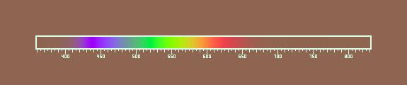
Visible spectrum (From Wikipedia,
the free encyclopedia)
White
light dispersed by
a prism into the colors of the optical
spectrum.
The visible
spectrum is the portion of the electromagnetic spectrum that is visible
to (can be detected by) the human eye. Electromagnetic radiation in this range
of wavelengths
is called visible light or simply light. A typical human eye will respond to wavelengths from about 390 to 750 nm.[1]
In terms of frequency, this corresponds to a band in the vicinity of 790–400 terahertz. A
light-adapted eye generally has its maximum sensitivity at around 555 nm (540 THz),
in the green
region of the optical spectrum (see: luminosity function). The spectrum does not,
however, contain all the colors that the human eyes and brain can distinguish. Unsaturated colors such as pink, or purple variations
such as magenta,
are absent, for example, because they can only be made by a mix of multiple
wavelengths.
Visible wavelengths also pass through the "optical window", the region of the
electromagnetic spectrum that passes largely unattenuated
through the Earth's atmosphere. Clean
air scatters
blue light more than wavelengths toward the red, which is why the mid-day sky
appears blue. The human eye's response is defined by subjective testing (see CIE), but atmospheric
windows are defined by physical measurement.
The
"visible window" is so called because it overlaps the human visible
response spectrum. The near infrared (NIR) windows lie just out of human
response window, and the Medium Wavelength IR (MWIR) and Long Wavelength or Far
Infrared (LWIR or FIR) are far beyond the human response region.
Many species can see
wavelengths that fall outside the "visible spectrum". Bees and many other insects can see
light in the ultraviolet, which helps them find nectar
in flowers.
Plant species that depend on insect pollination may owe reproductive success to
their appearance in ultraviolet light, rather than how colorful they appear to
us. Birds too can see into the ultraviolet (300–400 nm), and some have
sex-dependent markings on their plumage, which are only visible in the ultraviolet
range.[2][3]
History

Newton's
color circle, from Opticks of 1704, showing
the colors correlated with musical notes. The spectral colors from red to violet
are divided by the notes of the musical scale, starting at D. The circle
completes a full octave,
from D to D. Newton's circle places red, at one end of the spectrum, next to
violet, at the other. This reflects the fact that non-spectral purple colors are
observed when red and violet light are mixed.
Two of
the earliest explanations of the optical spectrum came from Isaac
Newton, when he wrote his Opticks, and from Goethe, in his Theory of Colours,
although earlier observations had been made by Roger Bacon
who first recognized the visible spectrum in a glass of water, four centuries
before Newton discovered that prisms could disassemble and reassemble white
light.[4]
Newton
first used the word spectrum (Latin for
"appearance" or "apparition") in print in
Newton
divided the spectrum into seven named colors: red, orange,
yellow, green, blue, indigo, and violet.
(Some schoolchildren memorize this order using the mnemonic ROY G. BIV.)
He chose seven colors out of a belief, derived from the ancient
Greek sophists,
that there was a connection between the colors, the musical notes, the known
objects in the solar system, and the days of the week.[5][6]
The human eye is relatively insensitive to indigo's frequencies, and some
otherwise well-sighted people cannot distinguish indigo from blue and violet.
For this reason some commentators, including Isaac
Asimov, have suggested that indigo should not be regarded as a color in its
own right but merely as a shade of blue or violet.
Johann Wolfgang von Goethe argued that
the continuous spectrum was a compound phenomenon. Where Newton narrowed the
beam of light to isolate the phenomenon, Goethe observed that a wider aperture
produces not a spectrum, but rather reddish-yellow and blue-cyan edges with white between them.
The spectrum only appears when these edges are close enough to overlap.
In the
early 19th century, the concept of the visible spectrum became more definite,
as light outside the visible range—ultraviolet
and infrared—was
discovered and characterized by William
Herschel, Johann Wilhelm Ritter, Thomas Young, Thomas Johann Seebeck,
and others.[7]
Young was the first to measure the wavelengths of different colors of light, in
1802.[8]
The
connection between the visible spectrum and color
vision was explored by Thomas Young and Hermann von Helmholtz in the early 19th
century. Their theory of color vision correctly proposed
that the eye uses three distinct receptors to perceive color.
Spectral colors
|
380–450 nm |
668–789 THz |
|
|
450–495 nm |
606–668 THz |
|
|
495–570 nm |
526–606 THz |
|
|
570–590 nm |
508–526 THz |
|
|
590–620 nm |
484–508 THz |
|
|
620–750 nm |
400–484 THz |
|
Colors
that can be produced by visible light of a single
wavelength (monochromatic light) are referred to as the pure
spectral colors.

Linear_visible_spectrum.svg
(SVG file, nominally 605 × 115 pixels, file size: 13 KB)
(http://upload.wikimedia.org/wikipedia/commons/d/d9/Linear_visible_spectrum.svg)
Although
the spectrum is continuous, with no clear boundaries between one color and the
next, the ranges may be used as an approximation.[9]
Spectroscopy
Rough
plot of Earth's atmospheric transmittance (or opacity)
to various wavelengths
of electromagnetic radiation, including
visible light.
Spectroscopy
is the study of objects based on the spectrum of color they emit or absorb.
Spectroscopy is an important investigative tool in astronomy
where scientists use it to analyze the properties of distant objects.
Typically, astronomical spectroscopy uses
high-dispersion diffraction gratings to observe spectra at very
high spectral resolutions. Helium was first detected by analyzing the spectrum of the Sun. Chemical
elements can be detected in astronomical objects by emission
lines and absorption lines. The shifting of spectral lines
can be used to measure the red shift or blue shift
of distant or fast-moving objects. The first exoplanets were discovered by
analyzing the Doppler shift of stars at a resolution that revealed
variations in radial velocity as small as a few meters
per second. The presence of planets was revealed by their gravitational
influence on the motion of the stars.
Color display spectrum
Color
spectrum generated in a display device.
Color
displays (e.g., computer monitors and televisions)
mix red, green, and blue color to create
colors within their respective color
triangles, and so can only approximately represent spectral colors, which
are in general outside any color triangle.

No
higher resolution available. Spectrum_(brown_background).png (575 ×
120 pixels, file size: 3 KB, MIME type: image/png)
(http://en.wikipedia.org/wiki/File:Rendered_Spectrum.png)
A
render of the color spectrum into the sRGB color
space on a brown background.
Colors
outside the color gamut of the display device result in negative values. If
color accurate reproduction of the spectrum is desired, negative values can be
avoided by rendering the spectra on a gray background. This gives an accurate
simulation of looking at a spectrum on a gray background.[10]
Scanning
The
world of desktop scanners has crossed the threshold of Deep Color
where scanners are capable of capturing a billion or more colors.
Part 3:
Flicker Sensitivity of the Chicken
Ref.:
Flicker:. اضطرب, تردد, رمش
Figures
See also
Definition of Contrast: Contrast is the difference in similarity and
comparison to others. It is emphasizing differences and showing distinctions as
in light versus darkness.
.png)
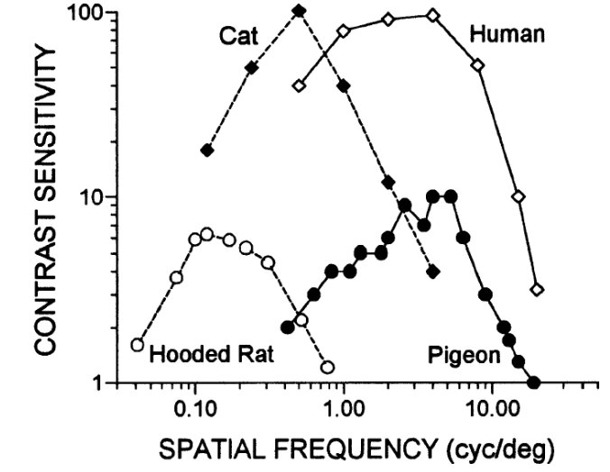
Figure - The contrast sensitivity function describes how
poor the contrast can become and still be perceptible. As the contrast
sensitivity value rises, the lower the contrast becomes. Combining the
experimental data with data for cats, pigeons, and humans, the hooded rat has
comparatively poor eyesight that may still be useful for medical studies (Ref.:
Vision in the Animal Kingdom: Vision in the Animal Kingdom (Kenneth Kang,
Psychology 221 - Applied Vision and Image Systems, Stanford University - Winter
2002).
In terms of the diversity of eyes and vision, birds
are quite amazing. While birds have a structure within their eye called a pecten whose purpose is not clear, they are capable of
seeing ultraviolet and polarized light with about four classes of cones. Their
cone cells have oil droplets to help distinguish colors. Moreover, they have a
higher flicker-fusion frequency, 100 Hz compared to 60 Hz for humans (Ref.:
Vision in the Animal Kingdom).
Ref.: (![]() )
)
References
1.
^ Cecie Starr (2005). Biology:
Concepts and Applications. Thomson Brooks/Cole. ISBN
053446226X. http://books.google.com/books?id=RtSpGV_Pl_0C&pg=PA94.
2.
^ Cuthill, Innes C; et al. (1997). "Ultraviolet
vision in birds". in Peter J.B. Slater. Advances in the Study of
Behavior. 29. Oxford, England: Academic Press. p. 161. ISBN
978-0-12-004529-7.
3.
^ Jamieson,
Barrie G. M. (2007). Reproductive Biology and Phylogeny of Birds.
Charlottesville VA: University of Virginia. p. 128. ISBN
1578083869.
4.
^ Coffey,
Peter (1912). The
Science of Logic: An Inquiry Into the Principles of Accurate Thought.
Longmans. http://books.google.com/books?id=j8BCAAAAIAAJ&pg=PA185&dq=%22roger+bacon%22+prism&ei=TX8OSJ2jMZCSzQTKx8y7Ag&client=firefox-a.
5.
^
Hutchison, Niels (2004). "Music For
Measure: On the 300th Anniversary of Newton's Opticks". Colour Music. http://home.vicnet.net.au/~colmusic/opticks3.htm.
Retrieved 2006-08-11.
6.
^ Newton,
Isaac (1704). Opticks.
7.
^ Mary Jo
Nye (editor) (2003). The
Cambridge History of Science: The Modern Physical and Mathematical Sciences.
5. Cambridge University Press. p. 278. ISBN
9780521571999. http://books.google.com/books?id=B3WvWhJTTX8C&pg=PA278&dq=spectrum+%22thomas+young%22+herschel+ritter&lr=&as_brr=0&as_pt=ALLTYPES&ei=XZT2Se_dF4vOkwT9tMigBA.
8.
^ John C.
D. Brand (1995). Lines
of light: the sources of dispersive spectroscopy, 1800-1930. CRC Press.
p. 30–32. ISBN
9782884491631. http://books.google.com/books?id=sKx0IBC22p4C&pg=PA30&dq=light+wavelength+color++young+fresnel&as_brr=3&ei=zpX2SdWLIpDmkASaxq3LBA#PPA31,M1.
9.
^ Thomas
J. Bruno, Paris D. N. Svoronos. CRC
Handbook of Fundamental Spectroscopic Correlation Charts. CRC Press,
2005.
10.
^ http://www.repairfaq.org/sam/repspec/
--------------------------
S45.4: UV vision and its functions in
birds
Innes C. Cuthill,
Julian C. Partridge & Andrew T. D. Bennett
School of Biological Sciences,
University of Bristol, Woodland Road, Bristol BS8 1UG, UK, fax 44 117 925 7374,
e-mail I.Cuthill@bris.ac.uk; J.C.Partridge@bris.ac.uk; Andy.Bennett@bris.ac.uk
Cuthill, I.C., Partridge, J.C. &
Bennett, A.T.D. 1999. UV vision and its functions in birds. In: Adams, N.J.
& Slotow, R.H. (eds)
Proc. 22 Int. Ornithol. Congr.,
Durban: 2743-2758. Johannesburg: BirdLife South Africa.
Birds can see ultraviolet (UV) light because, unlike
humans, their lens, cornea and other ocular media transmit UV, and they possess
a retinal cone type which is maximally sensitive to violet or ultraviolet
light, depending on the species. As birds also have cones sensitive to ‘blue’,
‘green’ and ‘red’ light, they may have a tetrachromatic
colour vision system.
Full article: http://www.int-ornith-union.org/files/proceedings/durban/Symposium/S45/S45.4.htm
--------------------------
Ultraviolet (UV) light perception by birds: a review, J. Rajchard Faculty of Agriculture, University of South
Bohemia, Ceske Budejovice, Czech Republic.
Veterinarni Medicina,
54, 2009 (8): 351–359: (http://www.vri.cz/docs/vetmed/54-8-351.pdf)
The ability to perceive (observe) the near ultraviolet part
of the light spectrum (the wavelength 320–400 nm) has been detected in many bird species.
It is now known that avian ocular media do not absorb UV
light before it reaches the retina; thus
UV sensitivity in birds is possible. Birds have 4–5 types of single cone
photoreceptors, including one type sensitive to UV light (for comparison humans
have only three types of cone photoreceptors). Many birds (obviously the
majority of species, e.g., many
non-passerines) have a violet-sensitive single cone that is obviously sensitive
to UV wavelengths. Other species (e.g.,
some passerines) have a single cone that has maximum sensitivity to UV light.
The spectral sensitivity of domestic ducks (Anas platyrhynchos domesticus) and turkeys (Meleagris
gallopavo gallopavo)
was tested over a range of specified wavelengths, including UVA, between
326–694 nm in comparison with human spectral sensitivity (Barber et al., 2006).
The results showed that ducks and turkeys had similar spectral sensitivities
and could perceive UVA radiation. Turkeys were more sensitive to UVA than
ducks. The peak sensitivity was in the wavelengths between 544–577 nm, with
reduced sensitivity at 508–600 nm. Both bird species had a very different and
broader range of spectral sensitivity than humans.
--------------------------
information. http://hsc.csu.edu.au/biology/options/communication/2950/CommPart2.html
|
Type of animal |
Name of animal |
Part of electromagnetic
spectrum detected |
Wavelengths detected |
|
Vertebrate |
Human |
visible |
700-400 nm |
|
|
Rattlesnake |
infra-red and visible |
850-480 nm |
|
|
Japanese dace fish |
ultraviolet and visible |
as low as 360 nm |
|
Invertebrate |
Honeybee |
ultraviolet and visible |
700-300 nm |
|
|
Mantis shrimp |
ultraviolet and visible |
640-400 nm |
Table:
(http://anthonymbiotask3.wikispaces.com/Light+and+the+electromagnetic+spectrum) or
http://hsc.csu.edu.au/biology/options/communication/2950/CommPart2.html
|
Type of animal |
Name of animal |
Electromagnetic spectrum used |
Reasons |
|
Vertebrate |
Human |
visible |
Active during the day uses colour for perception of objects |
|
|
Rattlesnake |
infra-red and visible |
Active at night hunts in dark burrows |
|
|
Hummingbird |
visible |
Can detect flowers from over a kilometre away |
|
Invertebrate |
Honeybee |
ultraviolet and visible |
Can detect ultraviolet markings on flowers and uses polarised light for navigation |
|
|
Mantis shrimp |
ultraviolet and visible |
Can perceive many more colours and escape predation in the well lit waters were it lives |
--------------------------
Part 4: Color
vision:
From Wikipedia, the free encyclopedia (http://en.wikipedia.org/wiki/Color_vision)
Color vision is the
capacity of an organism or machine to distinguish objects based on the wavelengths
(or frequencies)
of the light they
reflect, emit, or transmit. The nervous system derives color by comparing the
responses to light from the several types of cone
photoreceptors in the eye. These cone photoreceptors are sensitive to
different portions of the visible
spectrum. For humans, the visible spectrum ranges approximately from 380 to
740 nm, and there are normally three types of cones. The visible range and
number of cone types differ between species.
A 'red'
apple does not emit red light.[1]
Rather, it simply absorbs all the frequencies of visible
light shining on it except for a group of frequencies that is perceived as
red, which are reflected. An apple is perceived to be red only because the human eye
can distinguish between different wavelengths. The advantage of color, which is
a quality constructed by the visual brain and not a property of objects as
such, is the better discrimination of surfaces allowed by this aspect of visual
processing. In some dichromatic substances (e.g. pumpkin
seed oil) the color hue
depends not only on the spectral properties of the substance, but also on its
concentration and the depth or thickness[2].
Wavelength and hue detection
Isaac
Newton discovered that white light splits into its component colors when
passed through a prism, but that if those bands of colored light pass through
another and rejoin, they make a white beam. The characteristic colors are, from
low to high frequency: red, orange, yellow, green, cyan, blue, violet.
Sufficient differences in frequency give rise to a difference in perceived hue (hue: is the
degree to which a stimulus can be described as similar to or different from
stimuli that are described as red, green, blue,
and yellow); the just noticeable difference in wavelength
varies from about 1 nm in the blue-green and yellow wavelengths,
to 10 nm and more in the red and blue. Though the eye can distinguish up to a
few hundred hues, when those pure spectral colors are mixed together or diluted
with white light, the number of distinguishable chromaticities can be quite high.
In very
low light levels, vision is scotopic, meaning mediated by rod cells,
and not detecting color differences; the rods are maximally sensitive to
wavelengths near 500 nm. In brighter light, such as daylight, vision is photopic,
in which case the cone cells of the retina mediate color perception, and
the rods are essentially saturated; in this region, the eye is most sensitive
to wavelengths near 555 nm. Between these regions is known as mesopic vision, in which case both rods and cones are
providing meaningful signal to the retinal ganglion cells. The shift in color
perception across these light levels gives rise to differences known as the Purkinje
effect.
The
perception of "white" is formed by the entire spectrum of visible
light, or by mixing colors of just a few wavelengths, such as red, green, and
blue, or even by mixing just a pair of complementary colors such as blue and yellow.[3]
Physiology of color perception

Normalized response spectra of human
cones, short (S), medium (M), and long (L) types, to monochromatic spectral
stimuli, with wavelength given in nanometers.

The same figures as above represented here
as a single curve in three (normalized cone response) dimensions

Single
color sensitivity diagram of the human eye.
Perception
of color is achieved in mammals through color receptors containing pigments with
different spectral sensitivities. In most primates
closely related to humans there are three types of color
receptors (known as cone cells). This confers trichromatic color
vision, so these primates, like humans, are known as trichromats. Many other primates and other mammals are dichromats, and many mammals have little or no color
vision. Indeed, "mammals with color vision are rare," with most
mammals having rod-dominated retinas, and some having pure-rod ones.[4]
The
cones are conventionally labeled according to the ordering of the wavelengths
of the peaks of their spectral sensitivities: short (S), medium (M),
and long (L) cone types, also sometimes referred to as blue, green, and red
cones. While the L cones are often referred to as the red receptors, microspectrophotometry has shown that their peak
sensitivity is in the greenish-yellow region of the spectrum. Similarly, the S-
and M-cones do not directly correspond to blue and green, although they
are often depicted as such (such as in the graph to the right). It is important
to note that the RGB color model is merely a convenient means for
representing color, and is not directly based on the types of cones in the
human eye.
The peak
response of human color receptors varies, even amongst individuals with
'normal' color vision;[5]
in non-human species this polymorphic variation is even greater, and it may
well be adaptive.[6]
Theories of color vision
Two
complementary theories of color vision are the trichromatic theory
and the opponent process theory. The trichromatic
theory, or Young–Helmholtz theory, proposed in the 19th
century by Thomas Young and Hermann von Helmholtz, as mentioned above,
states that the retina's three types of cones are preferentially sensitive to
blue, green, and red. Ewald Hering
proposed the opponent process theory in 1872.[7]
It states that the visual system interprets color in an antagonistic way: red
vs. green, blue vs. yellow, black vs. white. We now know both theories to be
correct, describing different stages in visual physiology.[8]
Cone cells in the human eye
|
Cone type |
Name |
Range |
|
|
S |
β |
400–500 nm |
420–440 nm |
|
M |
γ |
450–630 nm |
534–545 nm |
|
L |
ρ |
500–700 nm |
564–580 nm |
A range
of wavelengths of light stimulates each of these receptor types to varying
degrees. Yellowish-green light, for example, stimulates both L and M cones
equally strongly, but only stimulates S-cones weakly. Red light, on the other
hand, stimulates L cones much more than M cones, and S cones hardly at all;
blue-green light stimulates M cones more than L cones, and S cones a bit more
strongly, and is also the peak stimulant for rod cells; and blue light stimulates
almost exclusively S-cones. Violet light appears to stimulate both L and S
cones to some extent, but M cones very little, producing a sensation that is
somewhat similar to magenta. The brain combines the information from each type
of receptor to give rise to different perceptions of different wavelengths of
light.
The
pigments present in the L and M cones are encoded on the X chromosome;
defective encoding of these leads to the two most common forms of color
blindness. The OPN1LW gene, which codes for the pigment that responds to
yellowish light, is highly polymorphic (a recent study by Verrelli and Tishkoff found 85
variants in a sample of 236 men[11]),
so up to twenty percent of women[12]
have an extra type of color receptor, and thus a degree of tetrachromatic color vision.[13]
Variations in OPN1MW, which codes for the bluish-green pigment, appear to be
rare, and the observed variants have no effect on spectral sensitivity.
Color in the human brain

Visual
pathways in the human brain. The ventral
stream (purple) is important in color recognition. The dorsal
stream (green) is also shown. They originate from a common source in the visual
cortex.
Color
processing begins at a very early level in the visual system (even within the
retina) through initial color opponent mechanisms. Opponent mechanisms refer to
the opposing color effect of red-green, blue-yellow, and light-dark. Visual
information is then sent back via the optic nerve
to the optic chiasma: a point
where the two optic nerves meet and information from the temporal (contralateral) visual field crosses to the other side of
the brain. After the optic chiasma the visual fiber
tracts are referred to as the optic tracts, which enter the thalamus to
synapse at the lateral geniculate
nucleus (LGN). The LGN is segregated into six layers: two magnocellular (large cell) achromatic layers (M cells) and
four parvocellular (small cell) chromatic layers (P
cells). Within the LGN P-cell layers there are two chromatic opponent types:
red vs. green and blue vs. green/red.
After synapsing
at the LGN, the visual tract continues on back toward the primary visual
cortex (V1) located at the back of the brain within the occipital
lobe. Within V1 there is a distinct band (striation). This is also referred
to as "striate cortex", with other cortical visual regions referred
to collectively as "extrastriate cortex".
It is at this stage that color processing becomes much more complicated.
In V1
the simple three-color segregation begins to break down. Many cells in V1
respond to some parts of the spectrum better than others, but this "color
tuning" is often different depending on the adaptation state of the visual
system. A given cell that might respond best to long wavelength light if the
light is relatively bright might then become responsive to all wavelengths if
the stimulus is relatively dim. Because the color tuning of these cells is not
stable, some believe that a different, relatively small, population of neurons
in V1 is responsible for color vision. These specialized "color
cells" often have receptive fields that can compute local cone ratios.
Such "double-opponent" cells were initially described in the goldfish
retina by Nigel Daw;[14][15]
their existence in primates was suggested by David
H. Hubel and Torsten Wiesel and
subsequently proven by Bevil Conway.[16]
As Margaret Livingstone and David Hubel showed, double opponent cells are
clustered within localized regions of V1 called blobs, and are thought to come
in two flavors, red-green and blue-yellow.[17]
Red-green cells compare the relative amounts of red-green in one part of a
scene with the amount of red-green in an adjacent part of the scene, responding
best to local color contrast (red next to green). Modeling studies have shown
that double-opponent cells are ideal candidates for the neural machinery of color
constancy explained by Edwin H. Land in his retinex
theory.[18]

This
image (when viewed in full size, 1000 pixels wide) contains 1 milion pixels, each of a different color. The human eye can
distinguish about 10 million different colors.[19]
From the
V1 blobs, color information is sent to cells in the second visual area, V2. The
cells in V2 that are most strongly color tuned are clustered in the "thin
stripes" that, like the blobs in V1, stain for the enzyme cytochrome oxidase (separating
the thin stripes are interstripes and thick stripes,
which seem to be concerned with other visual information like motion and
high-resolution form). Neurons in V2 then synapse onto cells in the extended
V4. This area includes not only V4, but two other areas in the posterior
inferior temporal cortex, anterior to area V3, the dorsal posterior inferior
temporal cortex, and posterior TEO.[20][21]
(Area V4 was identified by Semir Zeki
to be exclusively dedicated to color, but this has since been shown not to be
the case.[22]
Color processing in the extended V4 occurs in millimeter-sized color modules
called globs.[20][21]
This is the first part of the brain in which color is processed in terms of the
full range of hues
found in color
space.[20][21]
Anatomical
studies have shown that neurons in extended V4 provide input to the inferior temporal
lobe . "IT" cortex is thought to integrate color information with
shape and form, although it has been difficult to define the appropriate
criteria for this claim. Despite this murkiness, it has been useful to
characterize this pathway (V1 > V2 > V4 > IT) as the ventral
stream or the "what pathway", distinguished from the dorsal
stream ("where pathway") that is thought to analyze motion, among
many other features.
In other animals
Many invertebrates
have color vision. Honey-
and bumblebees have trichromatic color vision,
which is insensitive to red but sensitive in ultraviolet. Papilio
butterflies possess six types of photoreceptors and may have pentachromatic vision.[23]
The most complex color vision system in animal kingdom has been found in stomatopods (such as the mantis shrimp) with up to 12
different spectral receptor types thought to work as multiple dichromatic
units.[24].
Vertebrate
animals such as tropical fish and birds sometimes have more complex color vision systems than
humans.[25]
In the latter example, tetrachromacy is achieved
through up to four cone types, depending on species. Brightly colored oil
droplets inside the cones shift or narrow the spectral sensitivity of the cell.
It has been suggested that it is likely that pigeons are pentachromats.
Reptiles
and amphibians also have four cone types (occasionally five), and probably see
at least the same number of colors that humans do, or perhaps more. In
addition, some nocturnal geckos have the capability of seeing
color in dim light[26].
In the
evolution of mammals, segments of color vision were lost, then for a few
species of primates, regained by gene-duplication. Eutherian mammals other than primates (for example,
dogs, cats, mammalian farm animals) generally have less-effective two-receptor
(dichromatic)
color perception systems, which distinguish blue, green, and yellow—but cannot
distinguish reds. The adaptation to see reds is particularly important for
primate mammals, since it leads to identification of fruits, and also newly
sprouting leaves, which are particularly nutritious.
However,
even among primates, full color vision differs between new-world and old-world
monkeys. Old-world primates, including monkeys and all apes, have vision
similar to humans. New World Monkeys may or may not have color
sensitivity at this level: in most species, males are dichromats,
and about 60% of females are trichromats, but the owl monkeys
are cone monochromats, and both sexes
of howler
monkeys are trichromats.[27][28][29][30]
Visual sensitivity differences between males and females in a single species is
due to the gene for yellow-green sensitive opsin
protein (which confers ability to differentiate red from green) residing on the
X sex chromosome.
Several marsupials
such as the fat-tailed dunnart (Sminthopsis crassicaudata)
have been shown to have trichromatic color vision[31].
Marine
mammals, adapted for low-light vision, have only a single cone type and are
thus monochromats.
References
1.
^ Wright,
W. D. (1967). The rays are not coloured: essays on
the science and vision and colour. Bristol: Hilger. ISBN 0-85274-068-9.
2.
^ Kreft S and Kreft M (2007)
Physicochemical and physiological basis of dichromatic color, Naturwissenschaften 94, 935-939. On-line
PDF
3.
^
"Eye, human." Encyclopædia
Britannica 2006 Ultimate Reference Suite DVD, 2009.
4.
^ Ali,
Mohamed Ather; Klyne, M.A.
(1985). Vision in Vertebrates. New York: Plenum Press. pp. 174-175.
ISBN 0-306-42065-1.
5.
^ Neitz J, Jacobs GH (1986). "Polymorphism
of the long-wavelength cone in normal human color vision". Nature
323 (6089): 623–5. doi:10.1038/323623a0. PMID 3773989. http://www.nature.com/nature/journal/v323/n6089/abs/323623a0.html.
6.
^ Jacobs
GH (January 1996). "Primate
photopigments and primate color vision". Proc.
Natl. Acad. Sci. U.S.A. 93 (2): 577–81. doi:10.1073/pnas.93.2.577. PMID 8570598.
7.
^ Hering,
Ewald (1872). "Zur
Lehre vom Lichtsinne". Sitzungsberichte
der Mathematisch–Naturwissenschaftliche Classe der Kaiserlichen Akademie der Wissenschaften
LXVI. Band (III Abtheilung). http://books.google.com/books?id=u5MCAAAAYAAJ&pg=PA5&lpg=PA5&dq=1872+hering+ewald+Zur+Lehre+vom+Lichtsinne.+Sitzungsberichte+der+kaiserlichen+Akademie+der+Wissenschaften.+Mathematisch%E2%80%93naturwissenschaftliche+Classe,&source=web&ots=fAdrz1yI8x&sig=99NSKb_P8-_QSDO1RTzt35QTRyk&hl=en.
8.
^ Ali,
M.A. & Klyne, M.A. (1985), p.168
9.
^ Wyszecki, Günther; Stiles, W.S.
(1982). Color Science: Concepts and Methods, Quantitative Data and Formulae
(2nd ed.). New York: Wiley Series in Pure and Applied Optics. ISBN 0-471-02106-7.
10.
^ R. W.
G. Hunt (2004). The Reproduction of Colour
(6th ed.). Chichester UK: Wiley–IS&T Series in
Imaging Science and Technology. pp. 11–2. ISBN 0-470-02425-9.
11.
^ Verrelli BC, Tishkoff SA
(September 2004). "Signatures
of selection and gene conversion associated with human color vision
variation". Am. J. Hum. Genet. 75 (3): 363–75. doi:10.1086/423287. PMID 15252758.
12.
^ [Caulfield
HJ (17 April 2006). "Biological
color vision inspires artificial color processing". SPIE Newsroom.
doi:10.1117/2.1200603.0099. http://www.spie.org/x8849.xml?highlight=x2410.
13.
^ Roth,
Mark (2006). "Some
women may see 100 million colors, thanks to their genes" Post-Gazette.com
14.
^ Nigel
W. Daw (17 November 1967). "Goldfish Retina:
Organization for Simultaneous Color Contrast". Science 158
(3803): 942–4. doi:10.1126/science.158.3803.942.
PMID 6054169.
15.
^ Bevil R. Conway (2002). Neural
Mechanisms of Color Vision: Double-Opponent Cells in the Visual Cortex.
Springer. ISBN 1402070926. http://books.google.com/books?id=pFodUlHfQmcC&pg=PR7&dq=goldfish+retina+by+Nigel-Daw&as_brr=3&ei=2AWqR764JI7-iAGh8vwE&sig=7vvLHGgrRP_QtPH6mjLuiqblglU.
16.
^ Conway
BR (15 April 2001). "Spatial
structure of cone inputs to color cells in alert macaque primary visual cortex
(V-1)". J. Neurosci. 21 (8):
2768–83. PMID 11306629. http://www.jneurosci.org/cgi/content/full/21/8/2768.
17.
^ John E.
Dowling (2001). Neurons
and Networks: An Introduction to Behavioral Neuroscience. Harvard
University Press. ISBN 0674004620. http://books.google.com/books?id=adeUwgfwdKwC&pg=PA376&dq=Margaret+Livingstone+David+Hubel+double+opponent+blobs&as_brr=3&ei=YQaqR9-lAY6CiQHm1cmnCg&sig=D3znxI88shgNd8onK0RAWEMh6zY.
18.
^ McCann,
M., ed. 1993. Edwin H. Land's Essays. Springfield, Va.: Society
for Imaging Science and Technology.
19.
^ Judd,
Deane B.; Wyszecki, Günter (1975). Color in
Business, Science and Industry. Wiley Series in Pure and Applied Optics (3rd
ed.). New York: Wiley-Interscience.
p. 388. ISBN 0471452122.
20.
^ a
b
c
Conway BR, Moeller S, Tsao DY. (2007). Specialized
color modules in macaque extrastriate cortex. Neuron.
56(3):560-73. PMID
17988638
21.
^ a
b
c
Conway BR, Tsao DY. (2009). Color-tuned neurons are
spatially clustered according to color preference within alert macaque
posterior inferior temporal cortex. Proc Natl Acad Sci U S A. 106:18035-18039. PMID 19805195
22.
^ John Allman and Steven W. Zucker
(1993). "On
cytochrome oxidase blobs in visual cortex". in Laurence Harris and
Michael Jenkin, editors. Spatial Vision in Humans
and Robots: The Proceedings of the 1991 York Conference. Cambridge
University Press. ISBN 0521430712. http://books.google.com/books?id=eWBiKaOCNIYC&pg=PA34&dq=v4+zeki+color&lr=&as_brr=3&ei=KBCqR7eGF4bQiwHpnZSoCg&sig=F_rbsAj3FD69wRMzWGhB1vK4RuQ.
23.
^ Arikawa K (November 2003). "Spectral
organization of the eye of a butterfly, Papilio". J. Comp. Physiol.
A Neuroethol. Sens. Neural. Behav.
Physiol. 189 (11): 791–800. doi:10.1007/s00359-003-0454-7.
PMID 14520495. http://www.springerlink.com/content/whjepqnhpulyeevk/.
24.
^ Cronin
TW, Marshall NJ (1989). "A
retina with at least ten spectral types of photoreceptors in a mantis
shrimp". Nature 339: 137–40. doi:10.1038/339137a0. http://www.nature.com/nature/journal/v339/n6220/abs/339137a0.html.
25.
^ Kelber A, Vorobyev M, Osorio D
(February 2003). "Animal
color vision—behavioural tests and physiological concepts". Biol Rev Camb Philos Soc 78 (1): 81–118. doi:10.1017/S1464793102005985.
PMID 12620062. http://www.blackwell-synergy.com/doi/abs/10.1017/S1464793102005985.
26.
^ Roth, Lina S. V.; Lundström,
Linda; Kelber, Almut; Kröger, Ronald H. H.; Unsbo,
Peter (March 30, 2009). "The
pupils and optical systems of gecko eyes". Journal of Vision 9
(3:27): 1–11. doi:10.1167/9.3.27. http://journalofvision.org/9/3/27/.
27.
^ Jacobs,
G. H., & Deegan, J. F. (2001). Photopigments and color vision in New World monkeys from
the family Atelidae. Proceedings of the Royal
Society of London, Series B, 268, 695-702.
28.
^ Jacobs,
G. H., Deegan, J. F., Neitz,
J., Crognale, M. A., & Neitz,
(1993). Photopigments and color vision in the
nocturnal monkey, Aotus. Vision Research,
33, 1773-1783
29.
^ Mollon, J. D., Bowmaker, J. K.,
& Jacobs, G. H. (1984). Variations of color vision in a New World primate
can be explained by polymorphism of retinal photopigments.
Proceedings of the Royal Society of London, Series B, 222,
373-399.
30.
^
Sternberg, Robert J. (2006): Cognitive Psychology. 4th Ed. Thomson Wadsworth.
31.
^ Arrese CA, Beazley LD, Neumeyer C
(March 2006). "Behavioural evidence for
marsupial trichromacy". Curr.
Biol. 16 (6): R193–4. doi:10.1016/j.cub.2006.02.036.
PMID 16546067.
--------------------------
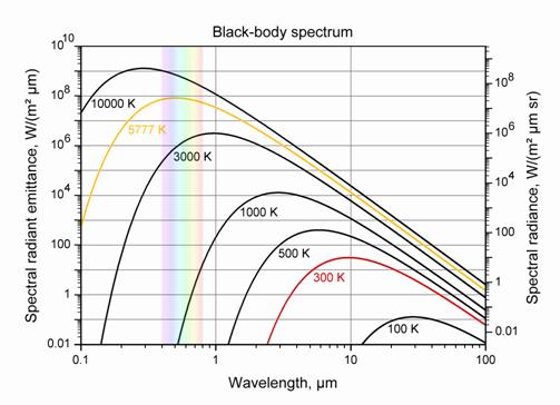
The color
temperature of a light source is the temperature
of an ideal black
body radiator that radiates light of comparable hue to that of the light
source. Color temperature is conventionally stated in the unit of absolute
temperature, the kelvin, having the unit symbol K.
To find the peak of the blackbody
radiation curve, Wien's Displacement Law gives: (http://hyperphysics.phy-astr.gsu.edu/hbase/wien.html)
![]()
If the temperature is 800K, then the
wavelength at which the radiation curve peaks is: λpeak
= 6225000000000005 microns
The corresponding frequency is
82815734989648.03313 Hz.
Planck's constant: h = 6.626068 × 10⁻³⁴
m2 kg / s.
The corresponding blackbody radiation
has photon energy
hν =
8.28157*6.626068*(10^-21)/(1.6*10^-19) = 0.342964 eV;
which is in the infrared range.
|
Name |
Wavelength |
Frequency (Hz) |
Photon Energy (eV) |
|
10 nm - 390 nm |
30 PHZ - 790 THz |
3 to 124 |
|
|
390 nm - 750 nm |
790 THz - 405 THz |
1.7 - 3.3 |
|
|
Infrared |
750 nm - 1 mm |
405 THz - 300 GHz |
0.00124 - 1.7 |
يقول الله سبحانه وتعالى: (خَلَقَ
الْإِنْسَانَ مِنْ صَلْصَالٍ كَالْفَخَّارِ وَخَلَقَ الْجَانَّ مِنْ مَارِجٍ مِنْ نَارٍ). إذا كانت درجة حرارة الْجَانَّ
تساوي أو أقل من 800K ، فان الأشعة الصادرة عنه تكون تحت الحمراء (hν = 0.342964 eV) فيتعذر على الإنسان رؤيته .
http://how-it-looks.blogspot.com/2010/01/infrared-radiation-black-bodies-and.html
Somewhere in the range 900K to 1000K,
the blackbody
spectrum encroaches enough in the the visible to
be seen as a dull red glow. Most of the radiated energy is in the infrared. (http://hyperphysics.phy-astr.gsu.edu/hbase/bbrc.html).
![]() To Arabic-English
To Arabic-English
![]() To English
To English